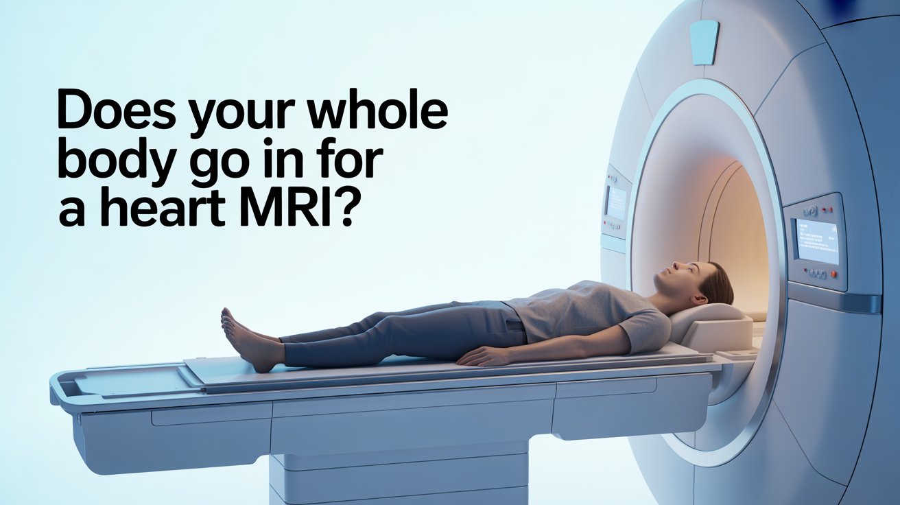
If you have been booked for a cardiac MRI and your biggest worry is whether you will be fully inside the scanner, the reassurance is this: you will not be. This is one of the most commonly searched questions before a cardiac MRI appointment — and the answer is simpler than most people expect.
A heart MRI focuses on the chest. That is the area the machine needs to image, and it is the area that enters the tunnel. Your head typically remains outside, and your legs and feet extend out the other end. The scanner is open at both ends throughout the entire scan.
If you are experiencing cardiac symptoms and want a prompt first assessment before being referred for advanced imaging, our ECG heart health check-up at The Private GP gives you same-day results with no waiting list.
Does Your Whole Body Go Into the Scanner?
No. For a cardiac MRI, only your chest and upper abdomen enter the tunnel. Your head stays outside at one end, and your legs and feet extend out the other.
Guy’s and St Thomas’ NHS Foundation Trust describes the MRI scanner as a large tube with a short, open tunnel through it. The tunnel is usually 60cm to 70cm wide and open at both ends. You lie on a bed that moves through the scanner — but for a cardiac scan, you slide in only far enough for your chest to be positioned within the imaging area.
Great Ormond Street Hospital NHS confirms that the MRI machine is shaped like a short, open-ended tunnel. The patient lies on a flat scanning bed that slides into the tunnel — but does not disappear entirely inside it.
For patients who are larger or broader across the shoulders, some centres offer a wider 80cm bore scanner. Guy’s and St Thomas’ advises patients who weigh more than 90kg to call ahead so the team can check which scanner is most appropriate for them.
What Happens During a Cardiac MRI?
You lie flat on a padded bed and are moved slowly into the scanner until your chest is positioned within the imaging area. The process is calm and methodical, and the radiographer communicates with you throughout via an intercom.
Guy’s and St Thomas’ NHS explains that ECG stickers are attached to your chest before the scan begins. These monitor your heartbeat in real time, allowing the machine to synchronise image capture with your heart rhythm — which is essential for producing clear, sharp cardiac images. If you have chest hair, a small area may need to be shaved to ensure the stickers adhere properly.
A blood pressure cuff may also be fitted to your arm and used to monitor your blood pressure at intervals during the scan.
What Does the Coil Do?
Before you enter the scanner, a flat pad called a coil is placed on your chest. Guy’s and St Thomas’ describes it as acting like an aerial for the scanner — it picks up the signals your body emits during imaging and converts them into higher quality pictures. A smaller pad called breathing bellows may also be placed on your abdomen to monitor your breathing pattern, which helps the machine capture certain images accurately.
You will be asked to hold your breath briefly at several points during the scan. The radiographer will tell you clearly when to hold and when to breathe normally. Most breath-holds last only a few seconds.
The scanner is noisy throughout — expect loud tapping and knocking sounds. Ear protection is always provided, and in most centres you can listen to music through headphones.
Will You Need Contrast Dye?
Not always. Contrast dye — a gadolinium-based agent injected through a small cannula in your arm — is used when the clinical team needs clearer images of blood flow, heart muscle scarring, or vessel detail. The British Heart Foundation explains that the dye makes images of blood flow to the heart show up more clearly on the scan.
If contrast is used, you will be advised to drink plenty of water afterwards to help flush the dye out of your system. If you have kidney problems, your doctor will check your kidney function before any contrast is given.
How Long Does a Heart MRI Take?
A standard cardiac MRI takes between 45 and 90 minutes, depending on the type of scan and the number of sequences needed.
University Hospital Southampton NHS confirms that you can expect your examination to take between 30 and 90 minutes depending on how complex your scan is. Guy’s and St Thomas’ advises allowing up to two hours for the full appointment, including preparation, the scan itself, and time afterwards if contrast or stress medication has been given.
A cardiac stress MRI — where medication is used to make your heart work harder, simulating the effect of exercise — takes longer than a standard scan, as two phases of imaging are required.
After the scan, a specialist analyses the images and prepares a written report, which is sent to the doctor who referred you.
What Conditions Can a Heart MRI Detect?
Cardiac MRI is one of the most comprehensive tests available for assessing the heart’s structure and function. It is often recommended when an echocardiogram (heart ultrasound) has not provided a complete enough picture.
The British Heart Foundation confirms that a cardiac MRI can help identify congenital heart disease, reduced blood flow to the heart muscle that may cause chest pain (angina), and other structural or functional abnormalities.
Conditions it can detect or help assess include:
Cardiomyopathy
Disease of the heart muscle — including hypertrophic, dilated, and arrhythmogenic types — where MRI shows the thickness, function, and scarring of the muscle in detail.
Heart attack scarring
Gadolinium contrast highlights areas of heart muscle damaged by a previous heart attack, distinguishing dead or scarred tissue from healthy muscle.
Congenital heart disease
Structural problems present from birth, such as holes between chambers or abnormal vessel connections, can be clearly mapped using cardiac MRI.
Heart valve disease
MRI can assess how well the valves are opening and closing and quantify the severity of any leakage or narrowing.
Pericardial disease
Inflammation or thickening of the sac surrounding the heart (the pericardium) shows up clearly on MRI.
Aortic abnormalities
Aneurysms, dissections, or narrowing of the aorta can be identified using MR angiography techniques within the same scan.
What Should You Know Before Your Cardiac MRI?
A few specific preparation steps apply to cardiac MRI that differ from other types of MRI.
Caffeine ban
Guy’s and St Thomas’ NHS advises avoiding all caffeine from midnight the night before your appointment. This includes coffee (regular and decaffeinated), tea, matcha, cola, and energy drinks. Caffeine raises the heart rate and interferes with image quality, particularly for stress perfusion scans.
Fasting
The American Heart Association advises that you may be asked not to eat or drink anything for four to six hours before the scan. Your appointment letter will confirm the exact requirement.
Metal and implants
Remove all jewellery, watches, and metal items before your appointment. If you have a pacemaker, implantable defibrillator, or any other metal device in your body, tell the cardiac MRI team as soon as possible. Many modern pacemakers are MRI-compatible, but this must be confirmed in advance.
Claustrophobia
If you are worried about the enclosed space, speak to your GP before the appointment. The American Heart Association confirms that a sedative can be prescribed to help you stay calm during the scan — but this must be arranged ahead of time. Some centres also offer wider bore scanners.
What to wear
Loose, comfortable clothing without metal zips or fasteners is ideal. A t-shirt and tracksuit bottoms or loose trousers are a practical choice.
If you are unsure whether a cardiac MRI is the right investigation for your symptoms, or if you need a referral, our private GP consultation can help you get the right answers quickly.
Frequently Asked Questions
- Will my head go inside the MRI machine for a heart scan?
For most cardiac MRIs, no. Your chest enters the tunnel but your head typically remains outside. The scanner is open at both ends throughout the scan.
- Is a cardiac MRI claustrophobic?
Some people find it uncomfortable, but most manage well. The scanner is open at both ends and your head stays outside. If you are anxious, a mild sedative can be prescribed by your GP before the appointment.
- Can I have a cardiac MRI if I have a pacemaker?
Many modern pacemakers are MRI-compatible, but this must be confirmed before your appointment. Always inform the cardiac MRI team about any implant as early as possible so they can check and plan accordingly.
- How long after a cardiac MRI will I get results?
A specialist analyses the images and sends a report to your referring doctor, typically within a few working days. Your doctor will then contact you to discuss the findings.
- Is a heart MRI better than an echocardiogram?
They serve different purposes. An echocardiogram is quicker and often the first test used. Cardiac MRI provides more detailed images of heart muscle, scarring, and complex structures — and is typically recommended when more information is needed beyond what the echocardiogram showed.