Most people know an ECG involves sticky pads and wires. But very few know what is actually happening in those 30 seconds while the machine is running — or what the clinician is looking at when they study the trace afterwards.
That uncertainty is one of the main reasons people feel anxious before the test. When you do not know what is happening, even a quick, painless procedure can feel daunting. The truth is, an ECG is one of the simplest and most elegant diagnostic tools in medicine. Once you understand what it is doing and why, the anxiety tends to disappear.
At The Private GP in Birmingham, our ECG heart health check-up is performed on site. You get results within minutes, discussed with you by a doctor who explains everything clearly. No jargon, no waiting, no uncertainty.
Here is exactly what happens — from the moment you walk in to the moment you walk out.
What Is an ECG Actually Doing to Your Heart?
An ECG does not do anything to your heart. It simply listens to it. Every time your heart beats, it produces a tiny electrical signal. The ECG machine detects those signals through electrodes on your skin and converts them into a visual trace that a clinician can read.
The American Heart Association explains that with each heartbeat, an electrical wave travels through the heart. This wave causes the muscle to squeeze and pump blood around the body. A normal heartbeat on an ECG shows the rate and rhythm of contractions in the upper and lower chambers of the heart.
Here is what that means in practice. Your heart has its own natural pacemaker, called the sinoatrial (SA) node, located in the upper right chamber. Every beat begins as a tiny electrical impulse fired from this node. That impulse travels through the upper chambers of the heart, causing them to contract and push blood downwards into the lower chambers. It then passes through a relay point called the atrioventricular (AV) node before spreading through the lower chambers, causing them to contract and push blood out to the lungs and body.
This entire electrical journey happens in under a second, with every single beat.
InformedHealth.org, published by NCBI, confirms that these electrical signals spread not just through the heart but throughout the body — all the way to the surface of the skin. That is how the electrodes on your wrists, ankles, and chest can pick them up without needing to go anywhere near the heart itself.
The ECG machine measures the changes in voltage on different areas of skin and plots them as a graph. That graph — with its distinctive peaks and dips — is your ECG trace.
What Happens Step by Step During an ECG?
A standard resting ECG follows a clear and predictable sequence. The whole thing, from entering the room to leaving again, takes around 5 to 10 minutes. The recording itself lasts less than a minute.
Here is exactly what you can expect.
You are taken to a private room
The clinician introduces themselves and briefly explains what the test involves. The NHS confirms you can request a chaperone — an additional member of staff to be present — at any point. Just ask if it has not been offered and you would like one.
You remove your upper clothing
You will be offered a gown or drape so that your chest is accessible while the rest of you remains covered. Dignity and privacy are maintained throughout.
Your skin is prepared
The clinician cleans the electrode sites on your chest, wrists, and ankles with a mild alcohol wipe. This removes any oils or lotions that could interfere with the signal. If you have significant chest hair, a small area may need to be shaved to ensure good contact.
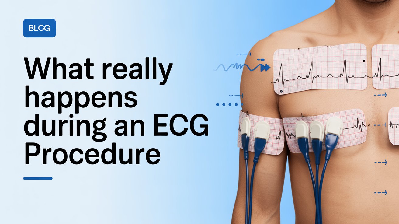
Ten electrodes are attached
The British Heart Foundation describes these as ten small sticky patches placed on your chest, arms, and legs. Six go across your chest in specific positions, and one goes on each wrist and ankle. Each electrode is connected by a wire to the ECG machine.
You lie still for the recording
The clinician presses a button to start the recording. You breathe normally and stay as still as possible. Moving, talking, or shivering can introduce interference into the trace. The recording itself is over in 30 to 60 seconds. You feel nothing at all during this time.
The electrodes are removed
Once the trace is complete, the wires are unclipped and the sticky patches are peeled away gently — similar to removing a plaster. The clinician will warn you before doing this.
You get dressed, and the results are reviewed
At The Private GP, your doctor reviews the trace immediately and discusses the findings with you during the same appointment. If you use our home visit service, the same process takes place in your own home.
What Are Those Squiggly Lines on the ECG Printout?
Each wave on the ECG trace represents a different part of a single heartbeat. The printout is, in effect, a story of your heart told beat by beat — and every peak and dip has a specific meaning that a trained clinician can read.
Research published via NCBI explains that the basic pattern of electrical activity across the heart was first identified over a hundred years ago. It comprises three main wave components: the P wave, the QRS complex, and the T wave. Together, they appear as a repeating pattern across the length of the trace — once for every heartbeat.
Here is what each part means in plain English.
The P wave is a small, rounded bump at the start of each cycle. It represents the electrical signal spreading across the upper chambers of the heart (the atria), causing them to contract and push blood downwards. If P waves are absent or irregular, it can suggest the heart is not generating rhythm from its usual starting point.
The QRS complex is the tall, sharp spike in the middle — the biggest and most recognisable feature of the trace. This represents the electrical signal spreading through the lower chambers (the ventricles), causing them to contract and push blood out to the rest of the body. It is the part of the trace most closely associated with each actual heartbeat.
The T wave is a broader, gentler wave that follows. It represents the ventricles resetting — what clinicians call repolarisation — ready to fire again on the next beat. Changes to the T wave’s shape, direction, or size can signal a range of conditions, from reduced blood flow to electrolyte imbalances.
A normal ECG trace shows these three components repeating in a steady, consistent pattern. The height of the waves, the spacing between them, and the shape of each peak all carry information. A clinician reading an ECG is essentially measuring the timing and pattern of your heart’s electrical circuit — beat by beat.
What Is the Clinician Actually Looking For?
When a doctor reviews your ECG trace, they are assessing several things simultaneously. Johns Hopkins Medicine confirms that an ECG records how fast the heart is beating, the rhythm of the heartbeats, and the timing of the electrical impulses as they move through the different parts of the heart.
In practice, the clinician is checking for:
Heart rate. A normal resting heart rate for adults is between 60 and 100 beats per minute. An ECG can identify both an unusually fast heart rate (tachycardia) and an unusually slow one (bradycardia) with precision.
Heart rhythm. The spacing between each QRS complex tells the clinician whether the heart is beating in a steady, regular pattern. Irregular spacing can indicate arrhythmias — abnormal heart rhythms — such as atrial fibrillation, which causes the upper chambers to quiver rather than contract properly.
Signs of a heart attack. Changes in the ST segment — the flat line between the QRS complex and the T wave — can indicate that part of the heart muscle is not receiving enough blood. An elevation of this segment is one of the key markers of a heart attack occurring right now. Changes in the pattern of Q waves can suggest a previous heart attack.
Ischaemia. This is the medical term for reduced blood flow to the heart muscle. It can show up as ST segment depression or changes to the T wave shape, suggesting that the heart’s blood supply is compromised even without a full heart attack.
Structural issues. Larger-than-expected wave amplitudes can suggest that certain chambers of the heart are enlarged or that the heart is working harder than it should be — sometimes a sign of high blood pressure, valve disease, or other conditions.
The British Heart Foundation is clear that an abnormal ECG reading does not always mean something is seriously wrong. Many findings require context — your symptoms, medical history, age, and other test results all form part of the picture. A single abnormal reading is the beginning of an investigation, not a definitive diagnosis.
Does an ECG Hurt, and Is It Safe?
An ECG is completely painless and carries no risk whatsoever. This is one of the most important things to understand before you have the test.
MedlinePlus, published by the US National Library of Medicine, is unambiguous: the machine does not send any electricity into your body. It only records electrical signals that your heart is already producing. There is no risk of electric shock, no radiation, and no invasive element of any kind.
The only sensation most people notice is when the sticky electrodes are peeled away at the end of the test. This is briefly uncomfortable — similar to removing a sticking plaster — particularly if you have chest hair. Occasionally, a mild rash or slight skin irritation may appear where the electrodes were placed, but this fades quickly.
Common concerns we hear from patients before their first ECG include:
“Will it hurt?” No. You feel nothing during the recording itself.
“Will I get an electric shock?” No. The machine only listens. Nothing is sent into your body.
“Will it affect my heart?” No. The ECG has no effect whatsoever on your heart’s function.
What Happens After the ECG Recording Is Complete?
Once the trace is printed, it is reviewed by a doctor. What happens next depends on where you have had the test.
At The Private GP, your results are reviewed on the same day — usually within minutes of the recording being taken. Your doctor goes through the trace with you, explains what it shows, and discusses any findings in plain language. You leave knowing exactly where your heart health stands.
If your ECG is normal, you will be given reassurance and, where relevant, advice about heart health monitoring going forward. We may discuss how frequently you should have a check, particularly if you have risk factors such as high blood pressure, high cholesterol, a family history of heart disease, or a history of smoking.
If something in the trace warrants closer investigation, this is not a reason to panic. Your doctor will explain what has been found, what it might mean, and what the next step is. This might include:
Private blood tests to check cardiac markers — for example, a BNP blood test, which measures a hormone released when the heart is under strain. This gives your doctor additional information about how hard the heart is working.
A repeat or extended ECG — either a second resting ECG or a 24-hour Holter monitor, which records your heart’s activity continuously while you go about your normal day.
A referral to a consultant cardiologist if specialist input is needed. We have strong referral networks and can arrange this promptly.
An ECG is often one part of a broader picture. Our full health check-up combines an ECG with blood pressure assessment, cholesterol testing, and other key health markers — giving you the most complete view of your cardiovascular health in a single appointment.
Frequently Asked Questions
- Will I feel anything during the ECG?
No. The recording is entirely painless. The only sensation most people notice is a mild tugging feeling when the sticky electrodes are removed at the end, similar to peeling off a plaster. Nothing is sent into your body during the test — the machine only listens to your heart’s existing electrical signals.
- Can I have an ECG if I have a pacemaker?
Yes. An ECG is safe to have with a pacemaker in place. Let your clinician know before the test begins, as the pacemaker’s signals will appear on the trace and need to be taken into account during interpretation. This is a routine consideration for trained clinicians and does not complicate the test significantly.
- What does it mean if the ECG trace is abnormal?
The British Heart Foundation is clear that an abnormal ECG does not automatically mean something is seriously wrong. Many findings are minor variations or require further context before a conclusion can be drawn. Your doctor will explain what has been found, what it might indicate, and what the appropriate next step is — whether that is a repeat test, blood tests, or a referral.
- Why do I need 10 electrodes if it is called a 12-lead ECG?
This is one of the most common questions we hear. InformedHealth.org explains that the standard 12-lead ECG uses 10 electrodes, but each electrode can be combined with others to create 12 different perspectives — or “leads” — of the heart’s electrical activity. Think of it as 12 different camera angles of the same event, captured using just 10 cameras.
- Can I eat and drink before an ECG?
Yes. The NHS confirms there is no need to fast before a standard resting ECG. You can eat and drink as normal and continue taking your usual medications. The one exception is if you are booked for an exercise ECG — in that case, your clinician will advise you to avoid a heavy meal and caffeine for a couple of hours beforehand.